
Product details
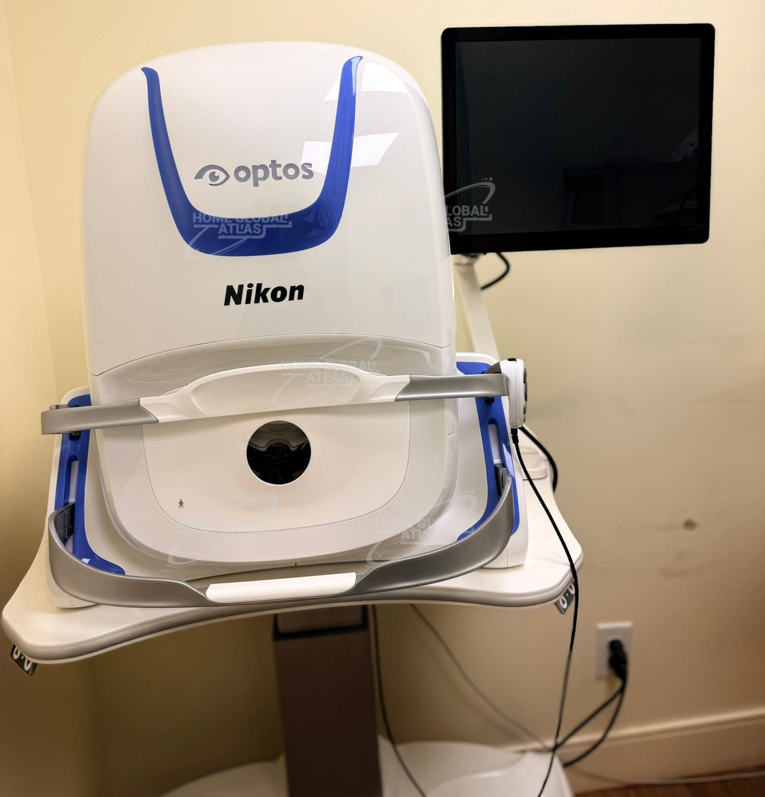
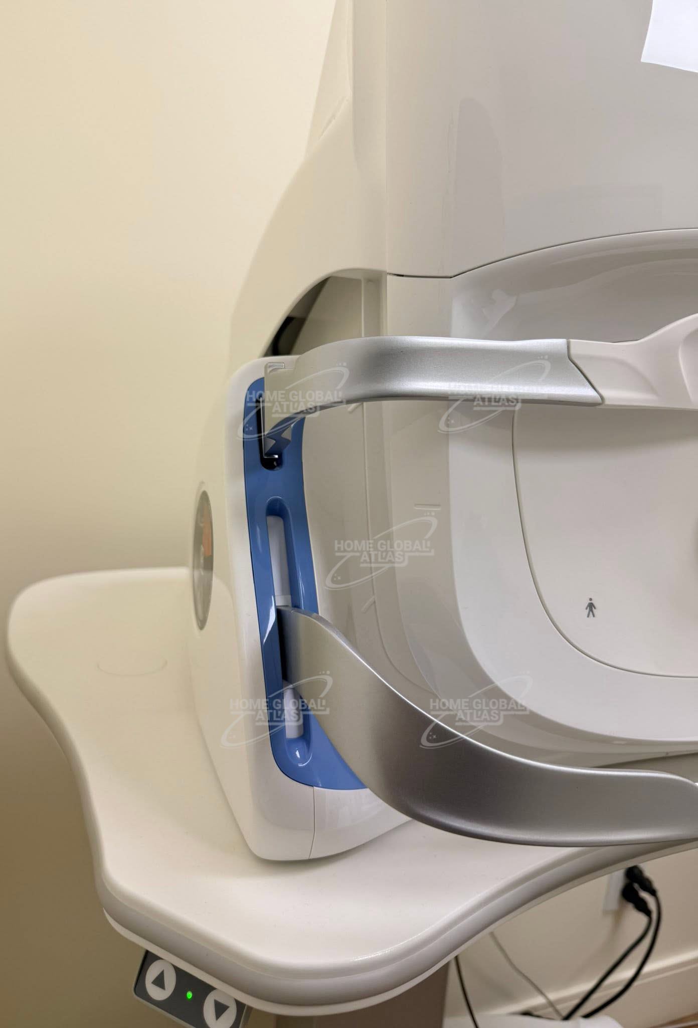
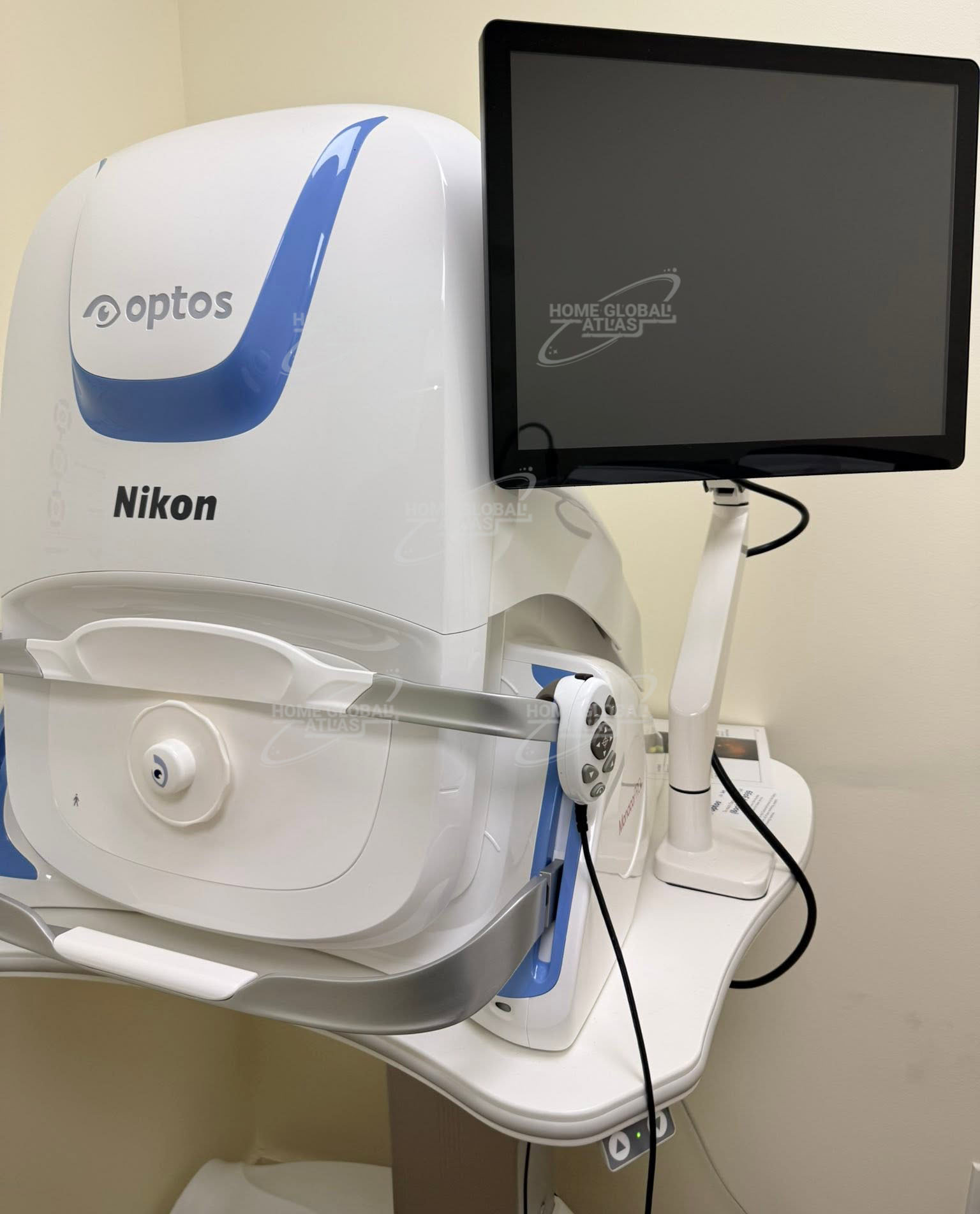
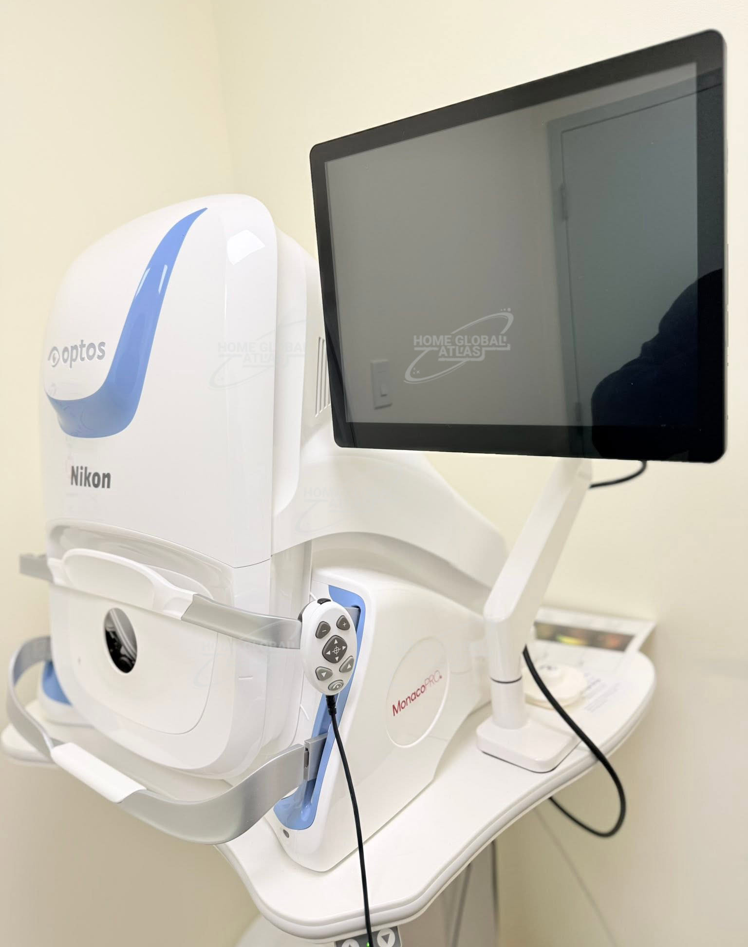
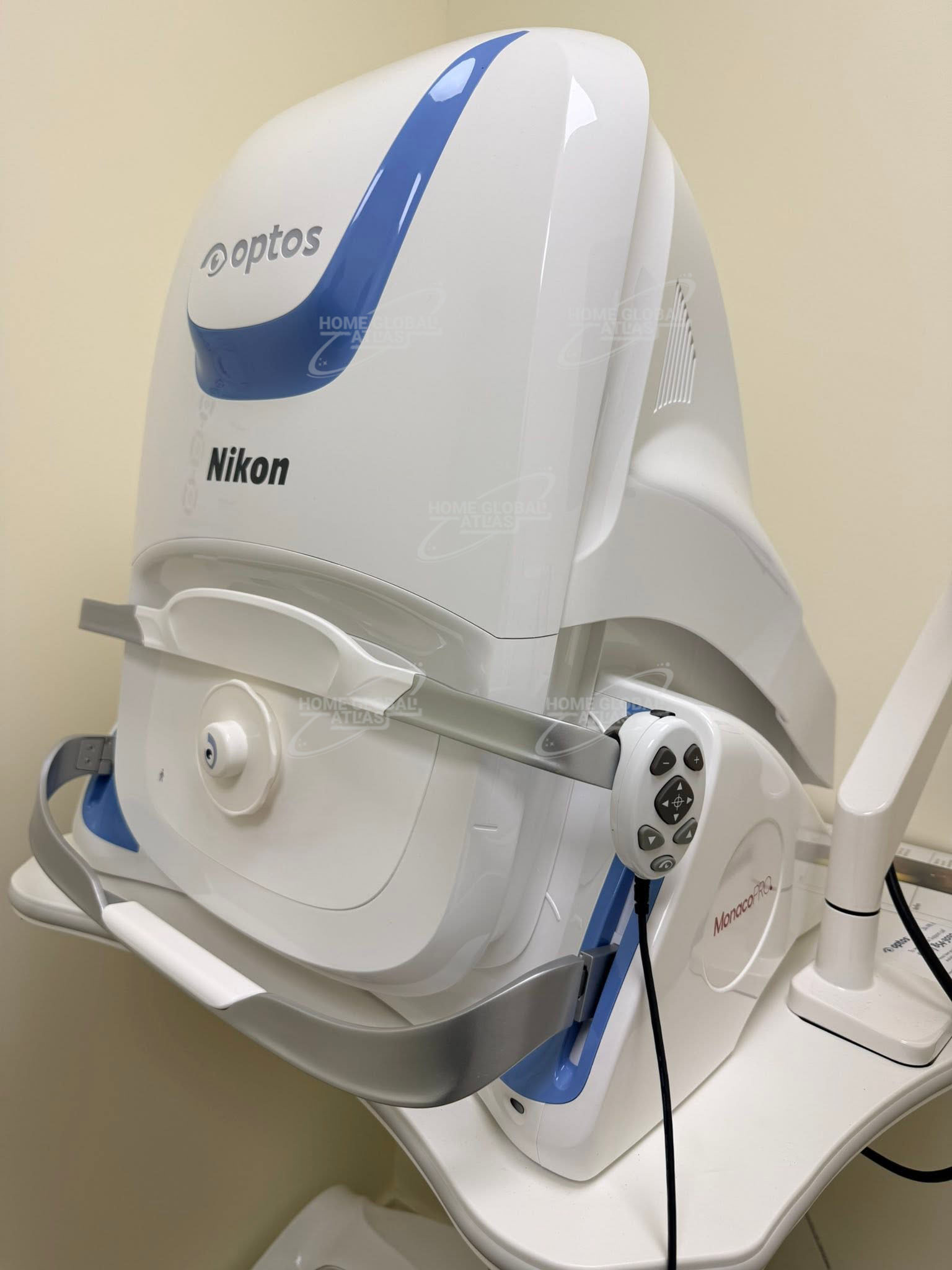
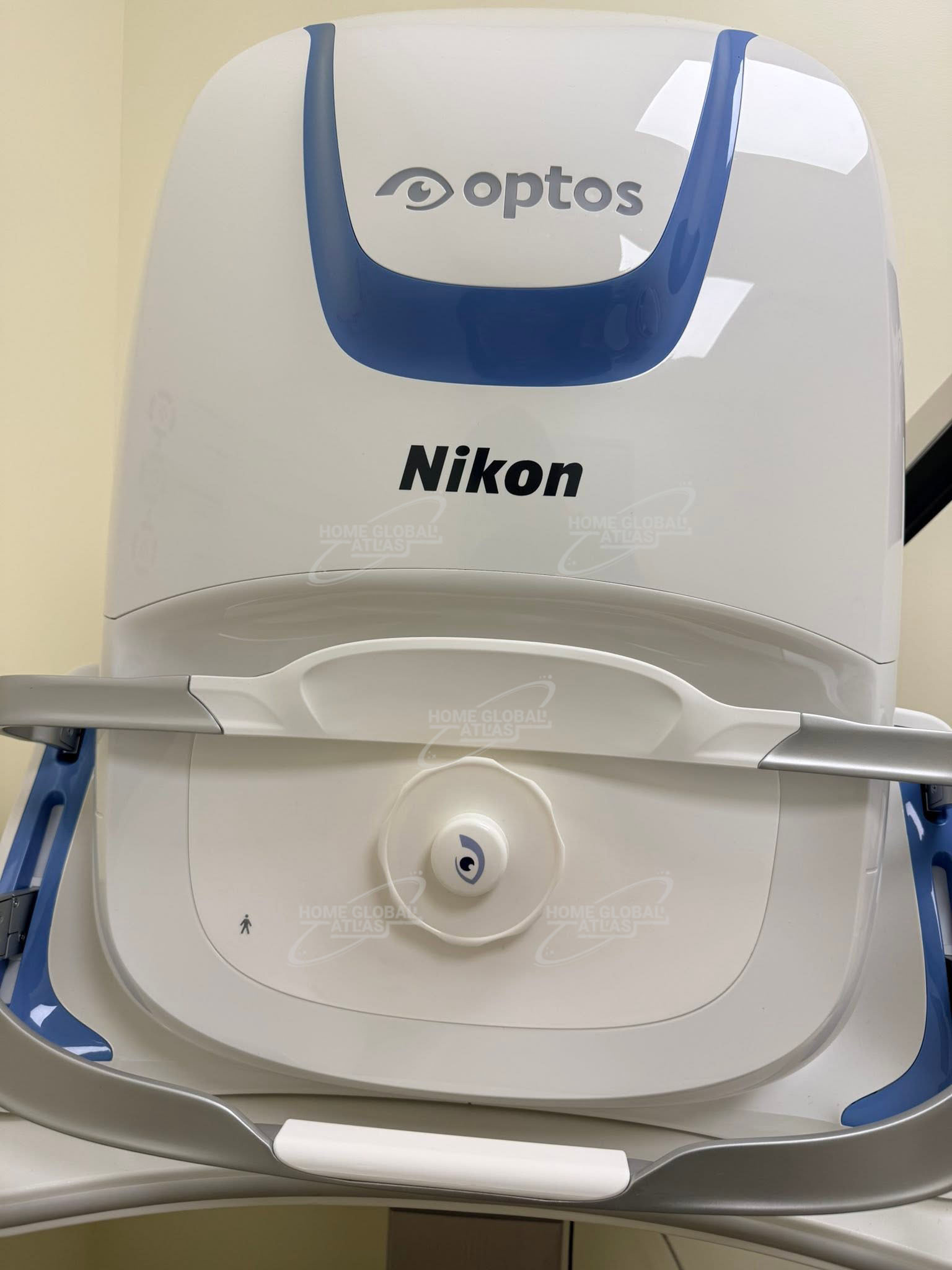
Optos Monaco Pro
Optos Monaco Pro is the sole UWF imaging device having an integrated OCT to help eye care professionals enhance their clinical examination and enhance practice economics.
Monaco combines optomap ultra-widefield technology with SD-OCT creating a fast, convenient, multi-modal imaging tool. Monaco can produce a 200°, single-capture retinal images of unrivaled clarity and can display a six-image overview including color, AF, and OCT of both eyes in as little as 90 seconds.
Monaco provides an exciting new way to enhance your clinical exam. The only ultra-widefield retinal imaging device with integrated OCT, Monaco produces a 200° single-capture optomap image in less than ½ second and also provides cross-sectional 40° OCT views of retinal structures. Monaco enables a rapid multi-modality capture featuring color, autofluorescence, and OCT scans, for both eyes, in as little as two minutes.
Home Global Atlas sell Optos Monaco Pro with best quality product
Monaco offers the following benefits:
• UWF with integrated OCT saves time, space and minimizes patient movement
• Central pole OCT provides comprehensive multi-modal imaging
• optomap images and OCT scans are correlated to facilitate pathology examination
• Color, AF, and OCT images are shown in a single, comprehensive view
Optos Monaco Retina Imaging Technical Specifications
Image Modalities
optomap color and optomap plus (red and green laser):
Color composite view
Green laser view
Red laser view
optomap af (green laser): autofluorescence
Optical Coherence Tomography (OCT)
Optomap Image Modalities
color rg
Sensory Retina
Choroidal
green af
SD-OCT
Resolution
optomap color: 20 μm
optomap plus, af : 14 μm
Exposure Time
Less than 0.4 seconds
OCT Scan Types Line Scan
Raster Scan
Retina Topography Scan
Optic nerve Head (OnH) Topography Scan
Retinal nerve Fiber layer (RnFl) Scan
Wavelengths Red laser: 635 nm
green laser: 532 nm (for af )
Tomographic Imaging
Signal Type: Optical scattering from tissue
Signal Source: Super luminescent Diode (SlD) 840 nm
Optical Power: laser safety Class-1 following IEC/en60825-1:2014
Typical Axial Resolution: <10 micron (in tissue) Digital on-screen <6 micron
Transverse Resolution: 20 micron (in tissue)
Scanners: Galavanometric with x, y mirrors
Scan Depth: Up to 2.5mm
OCT Scan Characteristics
Spectral Domain OCT
A-Scan rate up to 70k cycles/s
Active eye tracking
Automatic scan positioning
Footprint Width: 550 mm/22 inches
Depth: 570 mm/23 inches
Height: 606 – 632 mm/24-25 inches
There are no reviews yet.