
Product details
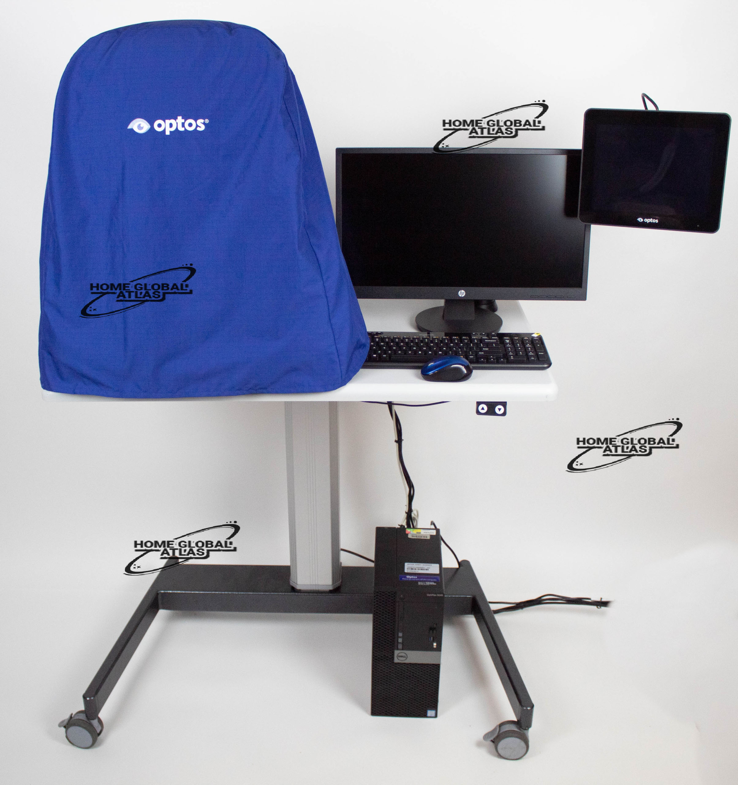
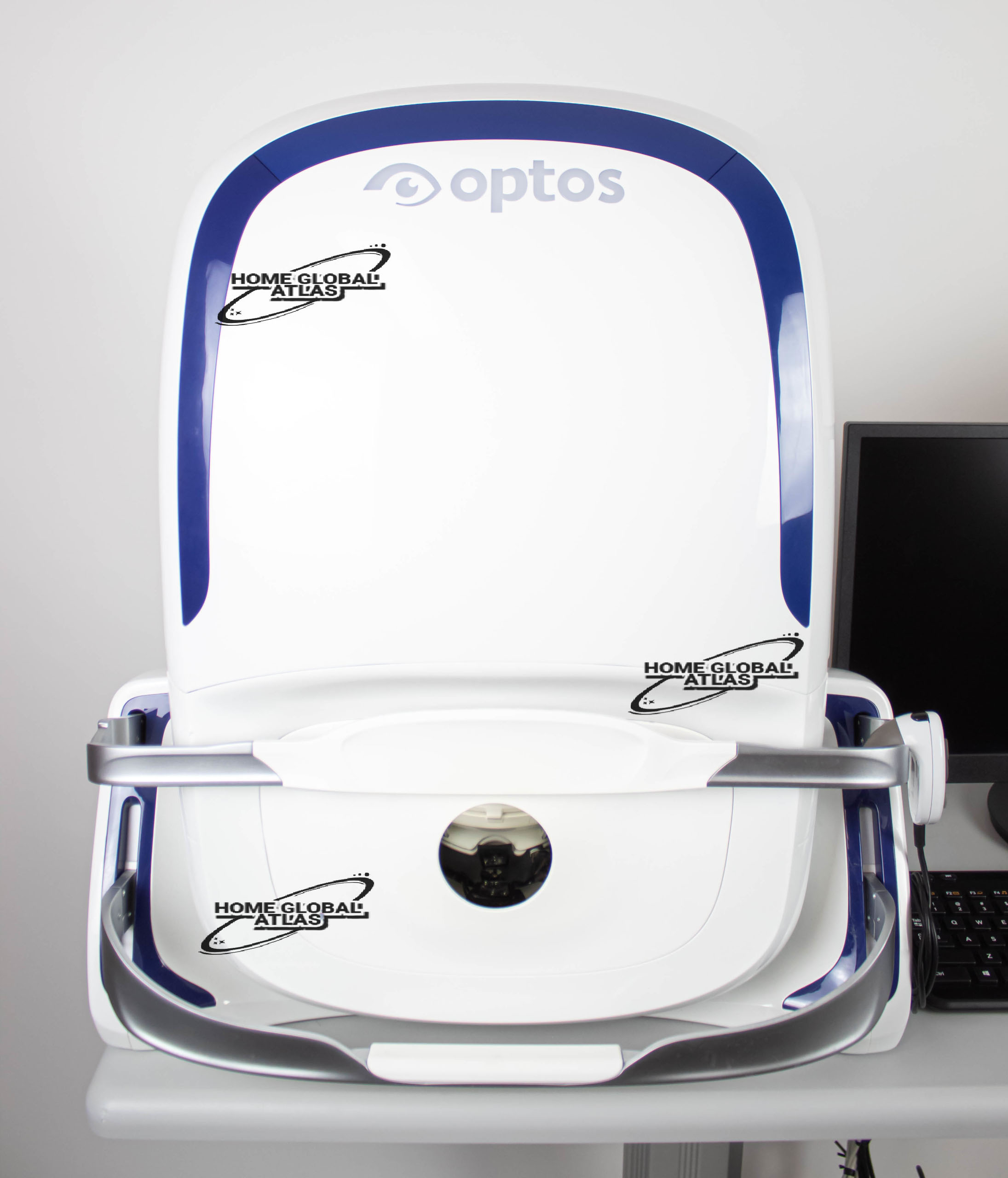
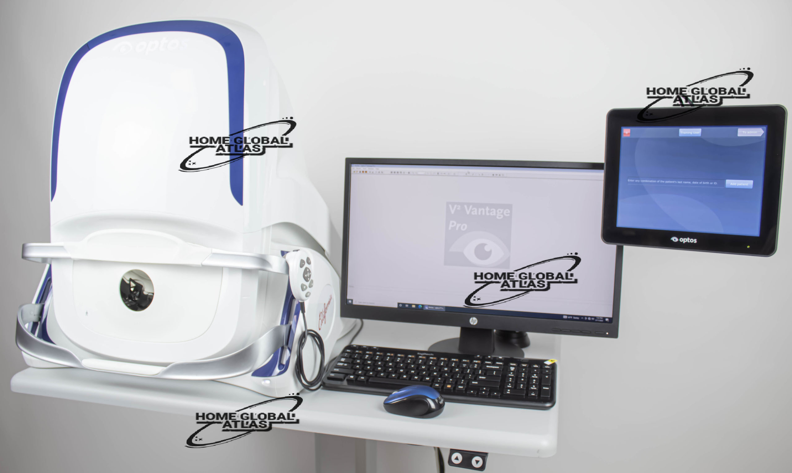
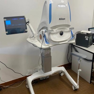
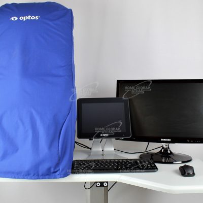
OPTOS California
OPTOS California is optomap imaging is proven to improve treatment decisions
optomap ultra-widefield (UWF) imaging is a proven tool for effective clinical decision making. More than 1,800 clinical trials and hundreds of published case studies and testimonials prove the value of optomap imaging in diagnosis, disease management, and patient engagement
Optos UWF technology is helping tens of thousands of eye doctors make better clinical decisions every day.
California was designed for medical imaging and is available in three models with multiple imaging modality options. California produces a 200°, single-capture retinal image of unrivaled clarity in less than ½ second and is changing management of diseases including DR, AMD, and uveitis.
With California, Optos has incorporated new hardware and software technology enabling practitioners to see more, discover more and effectively treat more ocular pathology thus promoting patient health.
In addition to the benefits found with all of the UWF devices from Optos such as 200 degrees or up to 82% of the retina captured in a single image, in multiple modalities, as well as eyecare professionals being able to see 50% more of the retina when compared to other conventional imaging devices, Californiarg, offers the following benefits:
– Motorized head and chin rest to more easily assist those patients requiring additional assistance during imaging
– Multiple imaging modalities including; 3-in-1 color depth imaging (color, red-free and choroidal in a single image) as well as
Autofluorescence
Home Global Atlas sell OPTOS California RG with best quality
OPTOS California RG Retina Imaging Features and Benefits:
Non-mydriatic imaging of the retina in less than ½ second saves time and helps improve clinic flow
cSLO technology images through most cataracts and small pupils (2 mm)
Multiple imaging modalities including; 3-in-1 color depth imaging (color, red-free and choroidal in a single image) as well as Autofluorescence provides important clinical data from the retinal surface through the choroid
Green laser autofluorescence shows macula and optic nerve head detail
Image overlay tool facilitates comparison of images in different image modes and from visit to visit
OptosAdvanceTM Image Management software streamlines image review and consultations
DICOM compatible software supports compliance with the Code of Federal Regulations 1,2
Distance (mm) and area (mm2) measurements provide objective assessment of change over time
Stereo disc imaging allows assessment of the optic nerve to diagnose and follow the progression of glaucoma
Auto-montage combines an optomap series into a single image showing up to 220° (97%) of the retina
Motorized head and chin rest to more easily assist those patients requiring additional assistance during imaging
Technical Specifications:
Image Modalities: optomap color and optomap plus (red and green laser):
Color composite view
Green laser view
Red laser view
OPTOS California RG Retina Imaging Technical Specifications
Image Modalities
optomap color and optomap plus (red and green laser):
Color composite view
Green laser view
Red laser view
optomap af (green laser): autofluorescence
Resolution
optomap color: 20 μm
optomap plus, af : 14 μm
Wavelengths
Red laser: 635 nm
Green laser: 532 nm
Exposure Time
Less than 0.4 seconds
Footprint
Width: 550 mm / 22 inches
Depth: 550 mm / 22 inches including chinrest
Height: 608-632 mm / 24-25 inches
Weight
34 kg/ 75 lbs
Laser Class
Laser safety Class-1 following IEC/EN60825-1:2007
and 21 CFR1040.10 and 1040.11
System Voltage
US: 100-120V at 50/60Hz
EU/AU: 200-240V at 50-60Hz
Power Consumption
Max. 300 VA
Communication Protocol
DICOM Compatible
optomap Image Modalities
color rgb (available with rg,af,fa combined model)
color rg
Sensory Retina
Choroidal
green af
blue af
fa
icg
There are no reviews yet.