
Product details
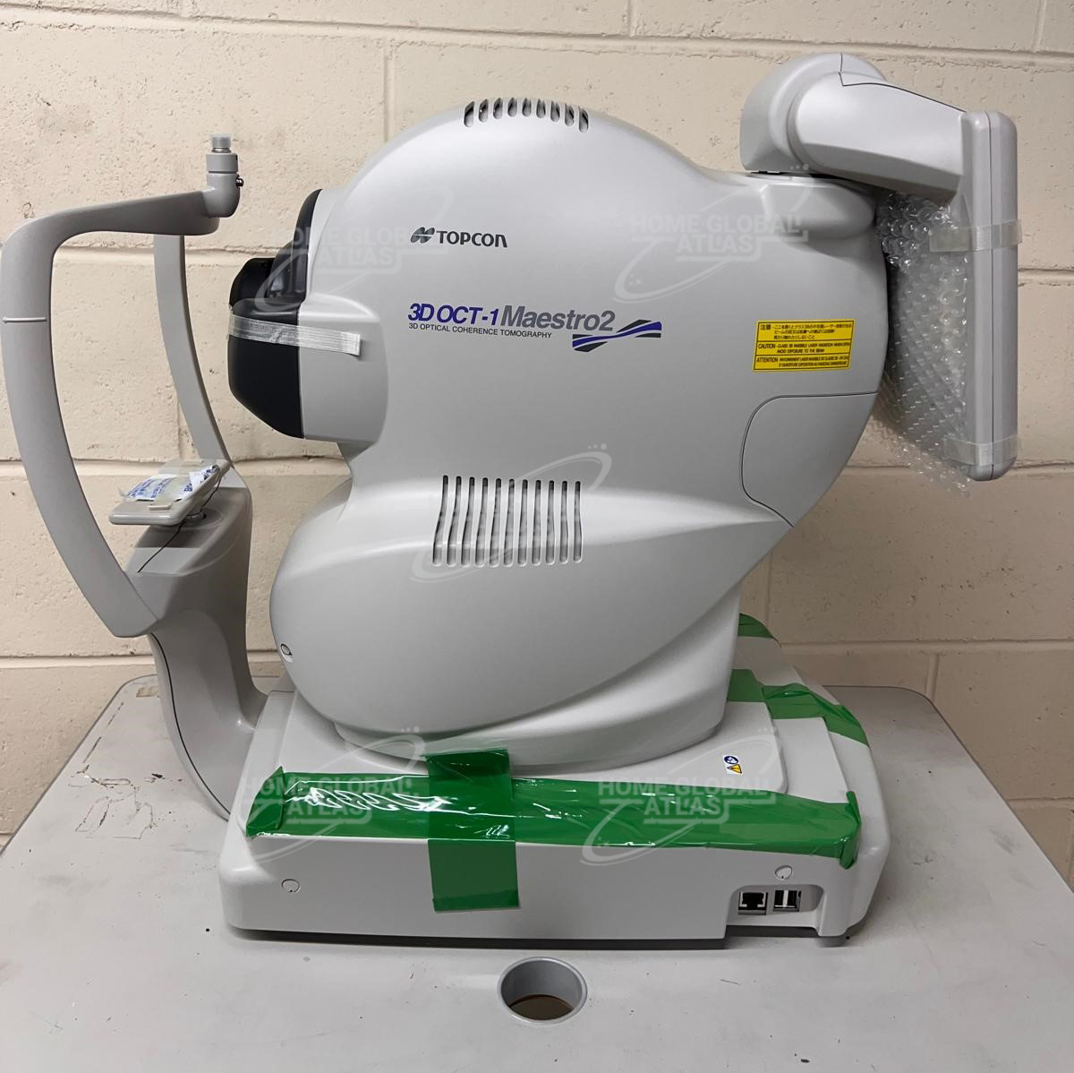
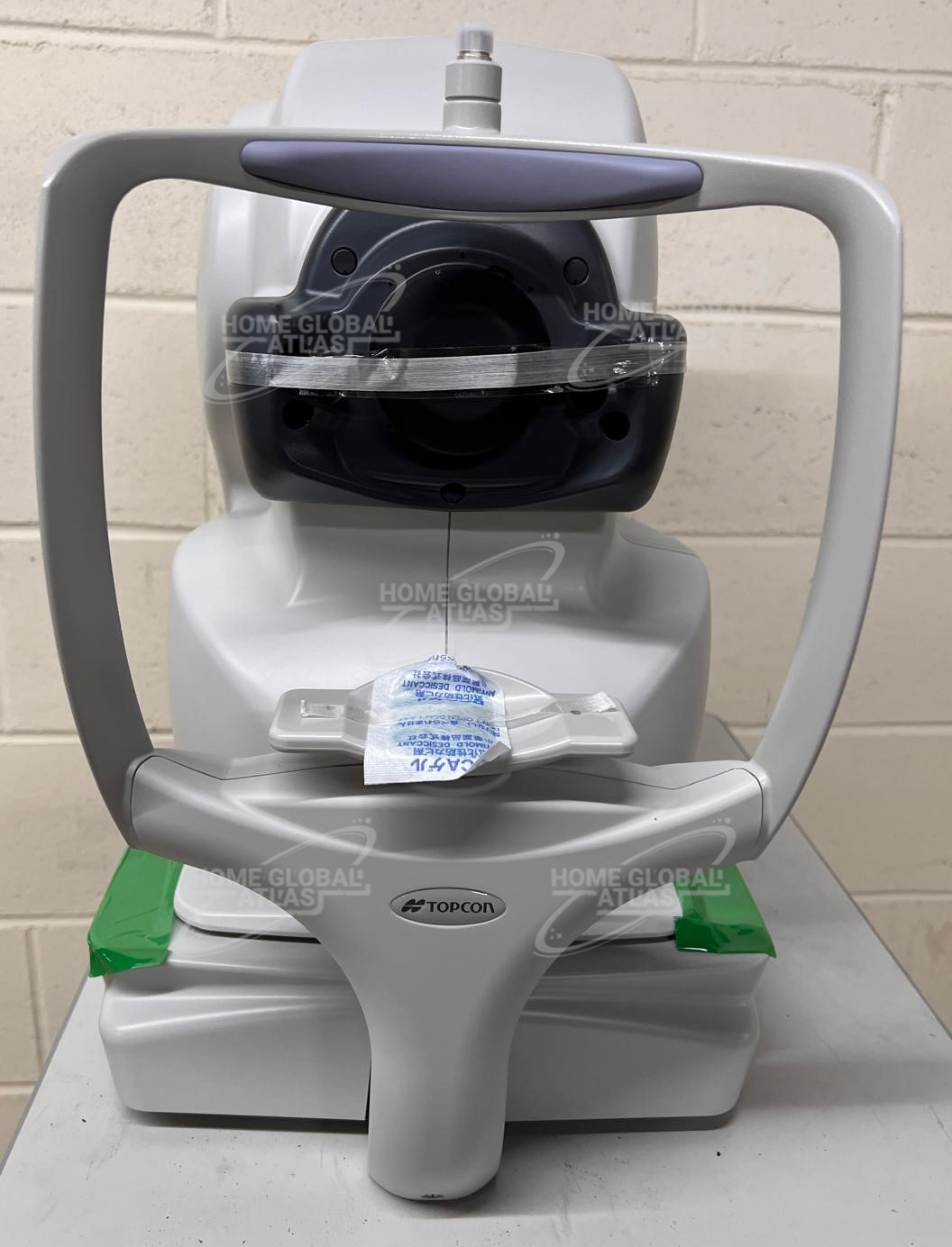
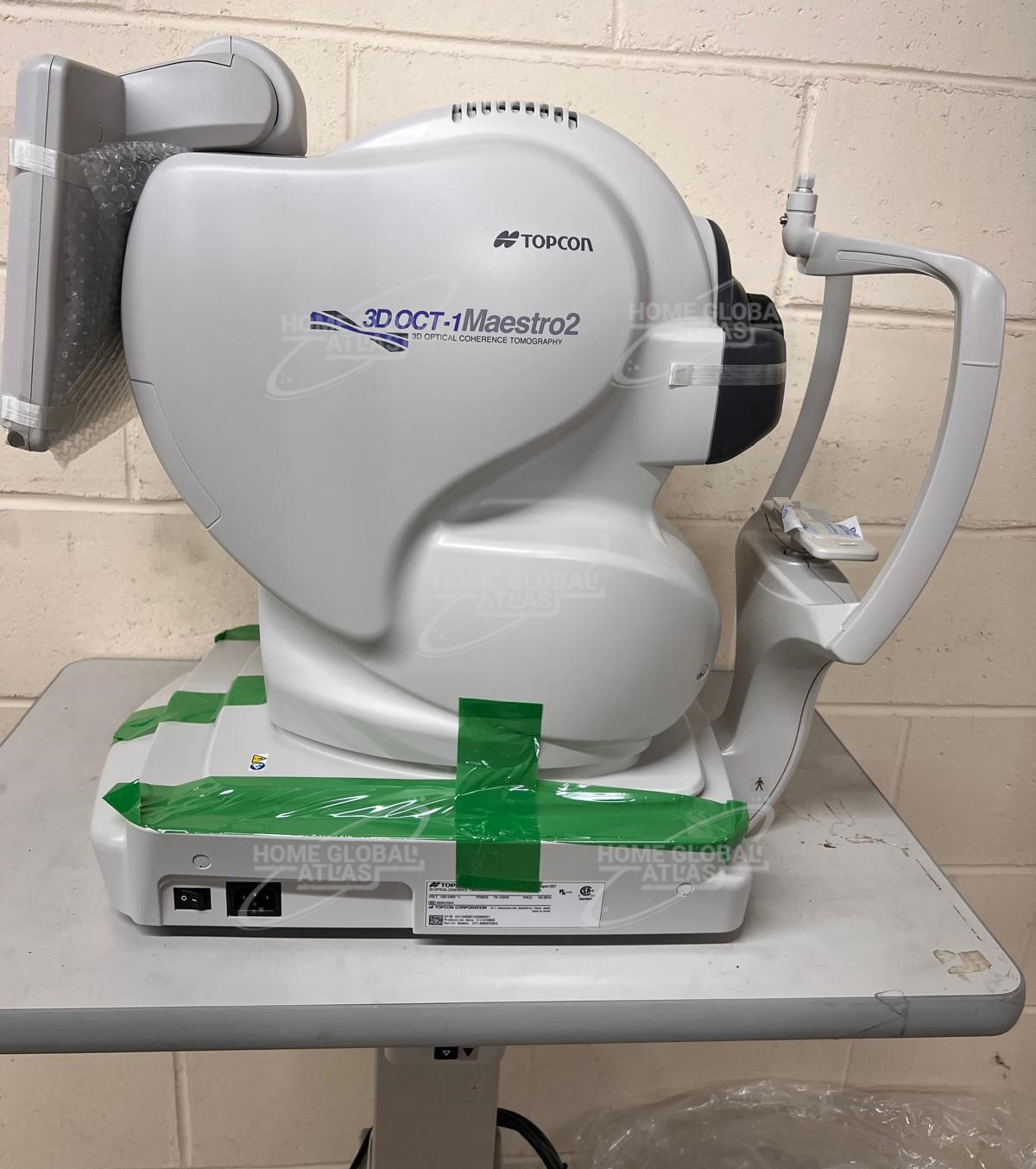
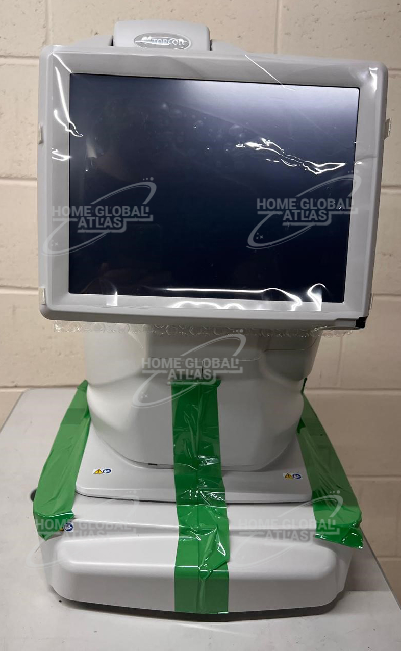
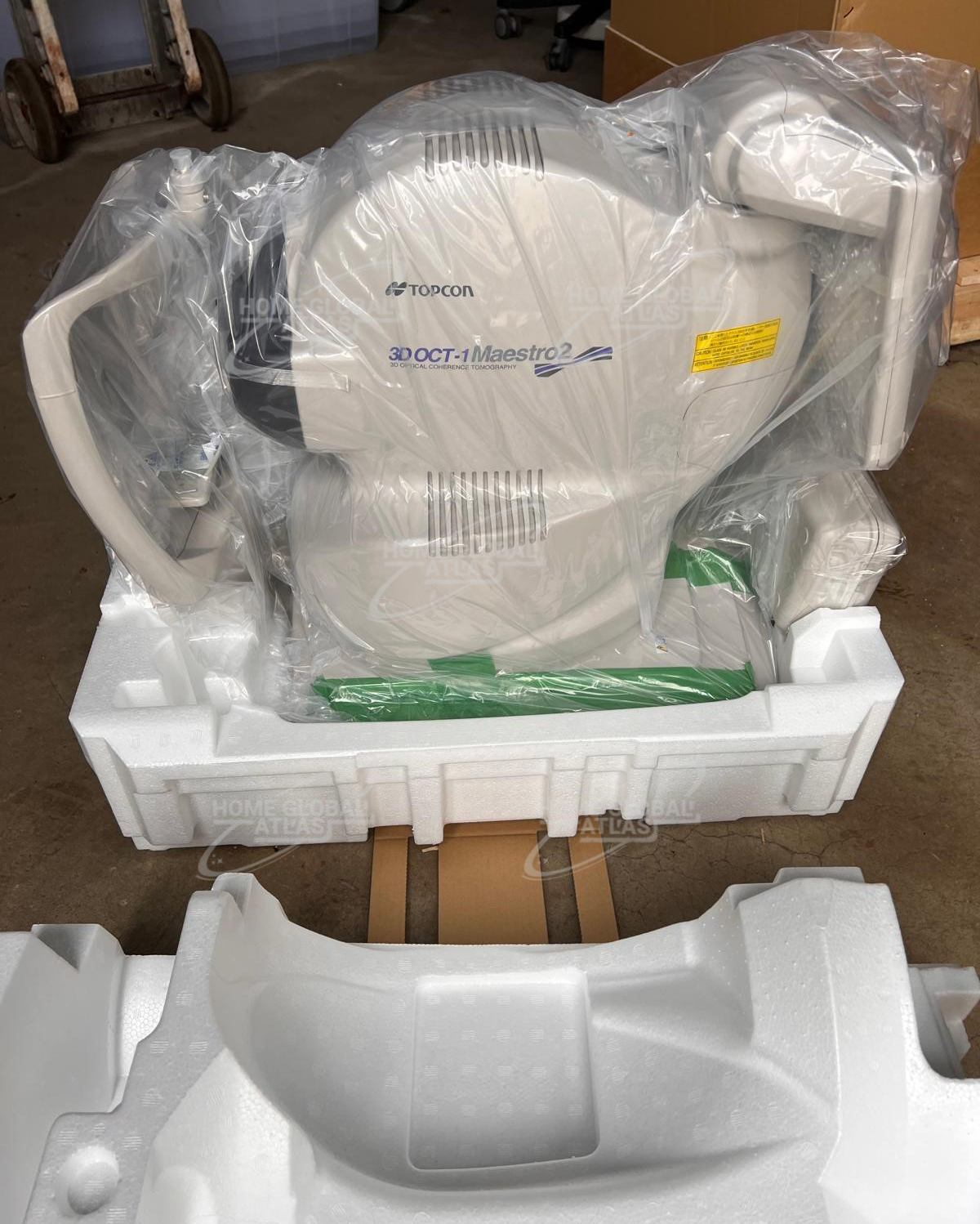
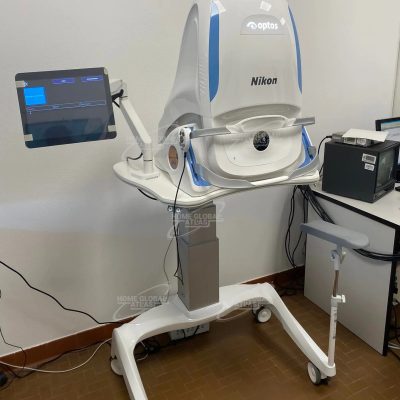
Topcon 3D OCT-1 Maestro 2
Topcon 3D OCT-1 Maestro 2 from ophthalmicmart provides ophthalmologists with an advanced automated optical coherence tomography system and fundus camera for assessing optical pathologies. Price Offer Available this Month Only.
With the Topcon Maestro2, you have fast, multi-modality OCT/Fundus imaging, and advanced data management. The Maestro2 is the complete clinical workstation for any busy practice. With a single touch, the Maestro2 automatically performs alignment, focus, optimizing and capturing. After capturing, the report can be immediately displayed by clicking on the icon. In addition to automated capture, the Maestro2 offers manual/semi-manual options for difficult-to-image patients.
Home Global Atlas sell Topcon 3D OCT-1 Maestro 2 with best quality product
The Maestro2 has an integrated full-color fundus camera. With one touch, you can simultaneously acquire a posterior OCT image and a true color fundus image. This allows for PinPoint Registration and structural confirmation of the pathology. A small pupil function is also available, as well as fundus only capture.
Topcon 3D OCT-1 Maestro 2 Key Features
Combination OCT and true color fundus
Compact and space-saving design
The NEW Hood Report for Glaucoma
Reference database for retina, RNFL, GCL+, and GCL++ thickness
Automatic layer segmentation
Widefield OCT
Anterior segment OCT
Panoramic fundus imaging
OBSERVATION & PHOTOGRAPHY OF FUNDUS:
Type of photography Color, Red-free* & IR
Picture angle of photography 45° ± 5% or less
30° or equivalent (digital zoom)
Operating distance 34.8 ± 0.1mm (when taking a picture of fundus)
Photographable diameter of pupil Normal pupil diameter: ø4.0mm or more
Small pupil diameter: ø3.3mm or more
Fundus image resolution (on fundus) Center: 60 lines/mm or more
Middle (r/2) : 40 lines/mm or more
Middle (r) : 25 lines/mm or more
IR photography : Center: 5 lines/mm or more***
OBSERVATION & PHOTOGRAPHY OF FUNDUS TOMOGRAM:
Scan range (on fundus) Horizontal direction 3 – 12mm ± 5% or less
Vertical direction 3 – 9mm ± 5% or less
Scan pattern 3D scan (horizontal/vertical)
Linear scan (Line-scan/Cross-scan/Radial-scan)
Scan speed 50,000 A-Scans per second
Lateral resolution 20µm or less
In-depth resolution 6µm or less
Photographable diameter of pupil ø2.5mm or more
OBSERVATION & PHOTOGRAPHY OF FUNDUS IMAGE / FUNDUS TOMOGRAM:
Fixation target Internal fixation target:
Dot-matrix type organic EL
The display position can be changed and adjusted.
The displaying method can be changed.
Peripheral fixation target:
This is displayed according to the internal fixation target displayed position.
External fixation target
OBSERVATION & PHOTOGRAPHY OF ANTERIOR SEGMENT:
Type of photography Color & IR
Operating distance 62.6 ± 0.1mm (when taking a picture of anterior segment)
OBSERVATION & PHOTOGRAPHY OF ANTERIOR SEGMENT TOMOGRAM:
Operating distance 62.6 ± 0.1mm (when taking a picture of anterior segment)
Scan range (on cornea)** Horizontal direction: 3 – 6mm ± 5% or less
Vertical direction: 3 – 6mm ± 5% or less
Scan pattern Linear scan (Line-scan/Radial-scan)
Scan speed 50,000 A-Scans per second
Fixation target External fixation target
There are no reviews yet.